XHSA-DCNet: An Explainable Hybrid Swin Transformer and Attention-Guided Dense Convolution Network for Automated Leukemia Detection and Classification
DOI:
https://doi.org/10.5281/zenodo.20541791Keywords:
Leukemia Detection, Medical Image Classification, Explainable AI, Deep Learning, Swin Transformer, Blood Smear ImagesAbstract
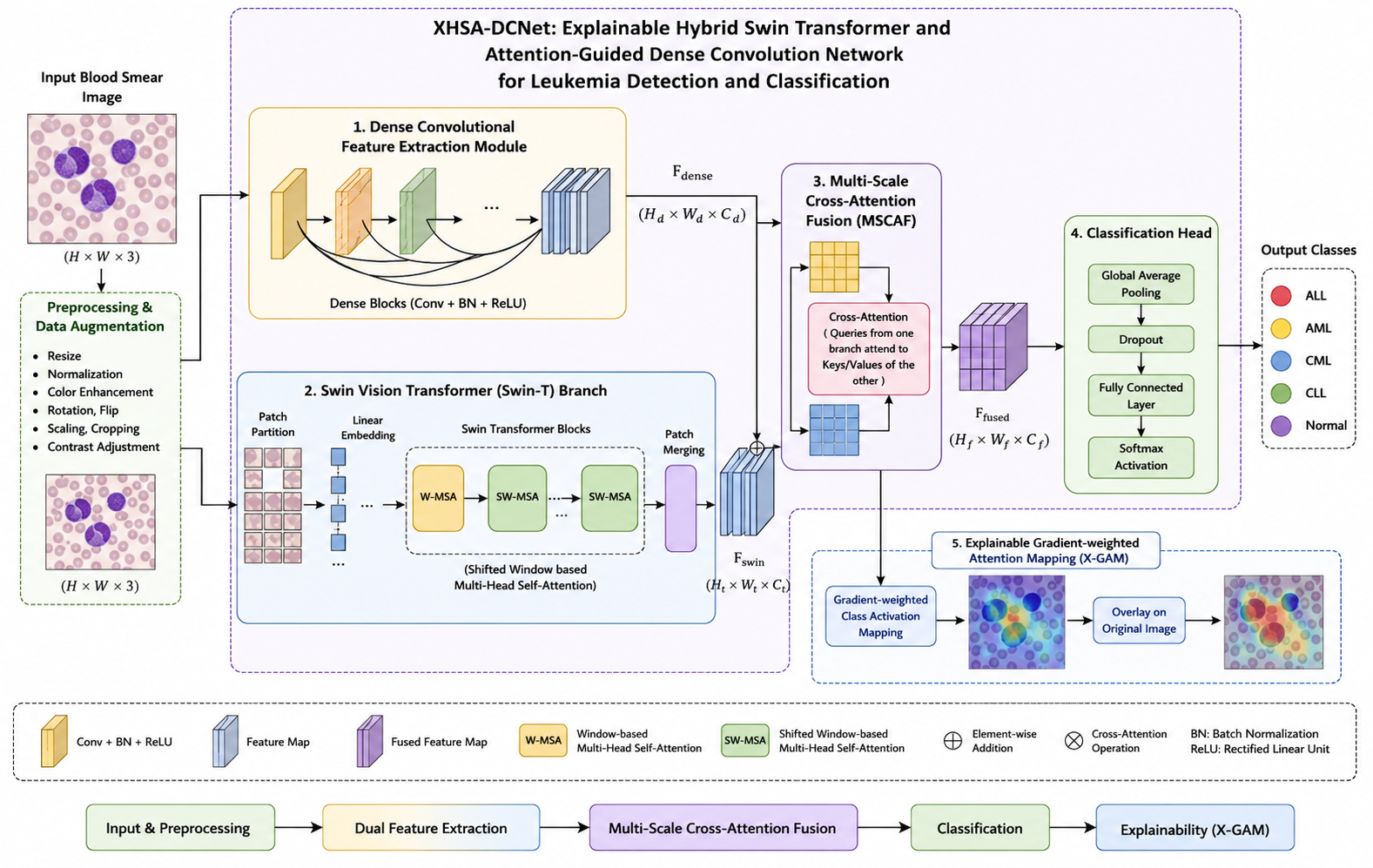
Leukaemia is a serious haematological cancer, where the abnormal growth of white blood cells needs to be diagnosed early and accurately, to ensure best treatment and survival. The manual microscopic analysis of blood smear images is a time consuming, labour intensive and skill intensive process which can only be performed by expert haematologists, which has encouraged the development of automated computer-aided blood smear diagnostic systems. This study aims to design an Explainable Hybrid Swin Transformer (XHSA-DCNet) for automatic detection and classification of leukemia from microscopic blood smear images, which combines explainable hybrid Swin Transformer and attention-guided dense convolution network. The suggested framework combines a Dense Convolutional Feature Extraction Module for extracting the fine-grained morphology of each cell and a Swin Vision Transformer for learning global contextual relationships. To effectively learn to fuse the local and global feature representations, a Multi-Scale Cross-Attention Fusion (MSCAF) module is introduced, and an Explainable Gradient-weighted Attention Mapping (X-GAM) mechanism to enhance model interpretability by highlighting diagnostically important regions of leukocytes. In addition, using advanced data augmentation and focal loss optimization for improving generalization and class imbalance problems. Experimental results show its effectiveness in terms of classification accuracy (99.1%), precision (98.9%), recall (99.0%), F1-score (98.95%) and AUC (0.995) outperforming CNN, ResNet50, DenseNet121, and Vision Transformer-based ones. The findings show that XHSA-DCNet is a promising clinical decision support system for haematologists and health care professionals, as it is highly accurate, robust and clinically interpretable for the diagnosis of leukemia.
References
[1] M. Loey, F. Smarandache, and N. E. M. Khalifa, “Within the lack of chest COVID-19 X-ray dataset: A novel detection model based on GAN and deep transfer learning,” Symmetry, vol. 12, no. 4, pp. 1–20, 2020.
[2] A. Ahmed, A. Nagy, A. Kamal, and D. Farghl, “Leukemia detection based on microscopic blood smear images using deep learning,” arXiv preprint arXiv:2301.03367, 2022.
[3] G. Vieira and M. E. Valle, “Acute Lymphoblastic Leukemia Detection Using Hypercomplex-Valued Convolutional Neural Networks,” arXiv preprint arXiv:2205.13273, 2022.
[4] F. M. Talaat and S. A. Gamel, “Machine learning in detection and classification of leukemia using C-NMC_Leukemia,” Multimedia Tools and Applications, vol. 83, pp. 8063–8076, 2024.
[5] H. H. R. Rasheed and A. M. Abdulazeez, “Leukemia Detection and Classification Based on Machine Learning and CNN: A Review,” Indonesian Journal of Computer Science, vol. 13, no. 3, pp. 1705–1721, 2024.
[6] F. Al-Obeidat et al., “Artificial intelligence for the detection of acute myeloid leukemia from microscopic blood images: A systematic review and meta-analysis,” Frontiers in Big Data, vol. 7, Art. no. 1402926, 2025.
[7] S. I. U. Rahman et al., “Deep Learning and Artificial Intelligence-Driven Advanced Methods for Acute Lymphoblastic Leukemia Identification and Classification: A Systematic Review,” Computer Modeling in Engineering & Sciences, vol. 142, no. 2, pp. 1199–1231, 2025.
[8] R. F. O. Kizi, T. P. T. Armand, and H.-C. Kim, “A Review of Deep Learning Techniques for Leukemia Cancer Classification Based on Blood Smear Images,” Applied Biosciences, vol. 4, no. 1, pp. 9–32, 2025.
[9] Aria, M., Javanmard, Z., Pishdad, D., Jannesari, V., Keshvari, M., Arastonejad, M., Safdari, R. and Akbari, M.E., “Towards Diagnostic Intelligent Systems in Leukemia Detection and Classification: A Systematic Review and Meta-analysis,” Journal of Evidence-Based Medicine, vol. 18, no. 1, e70005, 2025.
[10] Mollick, M. A. A., Rahman, M. M., Asadujjaman, D. M., Tamim, A., Dristi, N. A., & Hossen, M. T.., “Detection and Classification of Acute Lymphoblastic Leukemia Utilizing Deep Transfer Learning,” arXiv preprint arXiv:2501.14228, 2025.
[11] M. Maruf, M. M. Haque, and B. Paul, “Deep Learning with Self-Attention and Enhanced Preprocessing for Precise Diagnosis of Acute Lymphoblastic Leukemia from Bone Marrow Smears,” arXiv preprint arXiv:2508.17216, 2025.
[12] U. Ponnusamy and V. Perumal, “Comprehensive review on learning models of leukemia detection based on morphological information,” Leukemia & Lymphoma, vol. 67, no. 2, pp. 255–281, 2026.
[13] Shah, W.H., Fatima, S.R., Jaimes-Reátegui, R., Arévalo-Simental, D.E., Villalobos-Gutiérrez, P.T. and Pisarchik, A.N., “A systematic review of machine and deep learning techniques for acute lymphoblastic leukemia diagnosis,” Artificial Intelligence in Medicine, vol. 176, Art. no. 103393, 2026.
[14] Ghaderzadeh, M., Aria, M., Hosseini, A., Asadi, F., Bashash, D. and Abolghasemi, H., “A fast and efficient CNN model for B-ALL diagnosis and subtype classification using peripheral blood smear images,” International Journal of Intelligent Systems, vol. 37, no. 8, pp. 5113–5133, 2022.
[15] A. Mittal, S. Dhalla, S. Gupta, and A. Gupta, “Automated analysis of blood smear images for leukemia detection: A comprehensive review,” ACM Computing Surveys, vol. 54, no. 11, pp. 1–37, 2022.
Downloads
Published
Data Availability Statement
Data can be provided upon genuine request.
Issue
Section
License
Copyright (c) 2026 International Journal of Computational Intelligence in Engineering (IJCIE)

This work is licensed under a Creative Commons Attribution-ShareAlike 4.0 International License.